Surface analysis through spectral imaging
More fruitful than conventional RGB imaging, hyperspectral imaging allows a characterization of the optical properties of surfaces and materials beyond the single color attribute. The processing of the spectral content in the resulting images (IAU project), thanks to appropriate light scattering models (MMIS project), enables deducing therefrom, in each pixel, optical properties specific to the material under consideration. These methods have applications in the field of cosmetics (partnership with Newtone Technologies company), health as a diagnostic aid in dermatology (CHU Saint-Etienne), oncology and neurosurgery (LabEx Prime), and biology (partnership with BioMérieux) among others.
The light scattering models developped in this context find applications in many other fields, such as photovoltaics (optimisation of the optical properties of PV modules) and imaging systems (study of new generations of components, response of X-ray scintillators...).
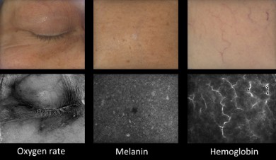
Example of information on human skin computed from hyperspectral images (400 - 700 nm in steps of 10 nm, 1 Mpx, acquisition time 2 s). Top row: color pictures rendered for a standard daylight illuminant. Bottom row: maps of skin component concentration estimated by a light scattering model (oxigen rate, melanin concentration, Hemoglobin concentration). Courtesy Newtone Technologies.
Seroul, P., Hébert, M., Cherel, M., Vernet, R., Clerc, R., & Jomier, M. (2016). Model-based skin pigment cartography by high-resolution hyperspectral imaging. Journal of Imaging Science and Technology, 60(6), 60404-1.